Dedifferentiated Chondrosarcoma of the Larynx: A Case Report and Review of the Literature- Juniper Publishers
Juniper Publishers- Journal of cell Science
Abstract
Background: Laryngeal chondrosarcomas are
rare, slow-growing, cartilaginous tumors. Dedifferentiated
chondrosarcomas, a rare entity of chondrosarcoma, are more aggressive
and associated with a more ominous prognosis. Definite diagnosis can be
established by incisional biopsy and histopathologic examination.
Histopathologic examination reveals a cartilaginous tumor with a
malignant spindle cell component. Definitive reatment of
dedifferentiated chondrosarcomas of the larynx is total laryngectomy.
There have been 14 case reports of laryngeal dedifferentiated
chondrosarcoma reported since 1988. The average life expectancy reported
is 6 months, and a 5-year survival rate of 10.5%.
Case presentation: We present a case of
dedifferentiated chondrosarcoma arising in the cricoids cartilage of a
male patient, who presented with 3-week history of dyspnea, stridor,
dysphonia and intermittent aphonia. As a result, he underwent a total
laryngectomy, and received adjuvant radiation therapy.
Conclusion: Laryngeal dedifferentiated
chondrosarcoma is a rare entity. Symptoms include dyspnea, hoarseness,
dysphagia, and a painless neck mass. Due to the aggressiveness of the
tumor, it is essential to include it as a differential diagnosis among
the other laryngeal tumors.
Keywords: Chondrosarcoma; Dedifferentiated; Larynx; Malignancy
Abbreviations:
DC: Dedifferentiated Chondrosarcoma; PL: Partial Laryngectomy; TL:
Total Laryngectomy; BND: Bilateral Neck Dissection; TT: Total
Thyroidectomy; WE: Wide Excision; ART: Adjuvant Radiotherapy; ARC:
Adjuvant Radiochemotherapy; DRMD: Died Of Regional Metastatic Disease;
DDMD: Died Of Distant Metastatic Disease; NED: Alive With No Evidence Of
Malignancy; AMD: Alive With Metastatic Disease; DOD: Died
Of Other Cause
Introduction
Chondrosarcoma (CS) is a slow-growing malignant
mesenchymal tumor with cartilaginous differentiation. CS, as a whole, is
relatively common [1-4].
These tumors are most commonly located in the pelvis, femur, ribs,
humerus, scapula, fibula, sacrum, or sternum; accounting for about
10-20% of malignant primary bone tumors [1-3,5-8].
Paranasal sinuses, nasal cavity, temporal bone, mandible and larynx
were the most common sites of head and neck chondrosarcomas origin [9]. Laryngeal chondrosarcomas are a raretumor, and account for only <0.2% of all head and neck malignancies [1,2,4,6,10-12].
Among the subtypes of CS, dedifferentiated CS (DC) is a rare, sinister
variant associated with a poor prognosis of an average life-expectancy
of 6 months, and a 5-year survival rate of 10.5%.
There have been numerous reports of laryngeal CS,
however, there are only 14 reports of laryngeal DC. Here we present a
case study of DC of the cricoid cartilage, including the clinical
presentation, investigations, and management.
Case Presentation
A 56-year-old Caucasian male was referred to the ENT
clinic with a 3-week history of dyspnea, stridor, dysphonia and
intermittent aphonia. Associated symptoms included frequent throat
clearing, globus pharyngeus, hoarseness and dysphagia. The patient lost
40 pounds over a 4- month period, and progressively became more
fatigued. He quit smoking 15 years ago. Prior to that, he smoked 10
cigarettes daily for 20 years. He denied the use of alcohol or
recreational drugs. Past medical history was significant for
hypertension, prediabetes, sleep apnea, COPD and GERD. The senior author
felt the later three diagnoses were secondary to the laryngeal
chondrosarcoma as they resolved post-laryngectomy. His head and neck
examinations were normal.
Flexible endoscopic laryngoscopy revealed left vocal
cord immobility, with the vocal cords in themedian position, and what
appeared endoscopically to be subglottic stenosis. He underwent a head,
neck and chest CT scan to rule out any sinister lesions along the course
of the recurrent laryngeal nerve. Imaging revealed a 4 x 4cm left
cricoid chondrosarcoma (Figure 1).

The CT scan revealed a chondrosarcoma, a poorly
defined mixed soft tissue and calcified mass measuring 3.0 x 2.0cm in
the axial dimension, and 3.5cm in the superior-inferior dimension,
centered on theposterior left lateral aspect of the cricoid cartilage.
The mass extended superiorly to the false vocal cords, and inferiorly,
just below the true vocal cords. Just below the true vocal cords, there
was significant narrowing of the airway. The lumen measured 2.5cm in the
anterior-posterior dimension, and only 4mm in the transverse dimension.
Destruction of most of the cricoid cartilage was identified. There was
no evidence of cervical lymphadenopathy. The lungs were clear and there
was no mediastinal lymphadenopathy. Diffuse fatty filtration of the
liver was present with a poorly defined 1.2cm hypodense mass in the
right lobe.
Shortly after the CT scan was done, the patient was
admitted to hospital with severe respiratory distress and complete
aphasia, he subsequently underwent a microlaryngoscopy, biopsy and
tracheostomy. The patient's dyspnea resolved following the procedure.
Due to the histological findings concluding laryngeal chondrosarcoma
with a non-functional larynx, he underwent a narrow field total
laryngectomy, cricopharyngeal myotomy, and tracheoesophageal puncture
with placement of voice prosthesis. Postoperatively, he recovered well
and his speech was 100% intelligible.
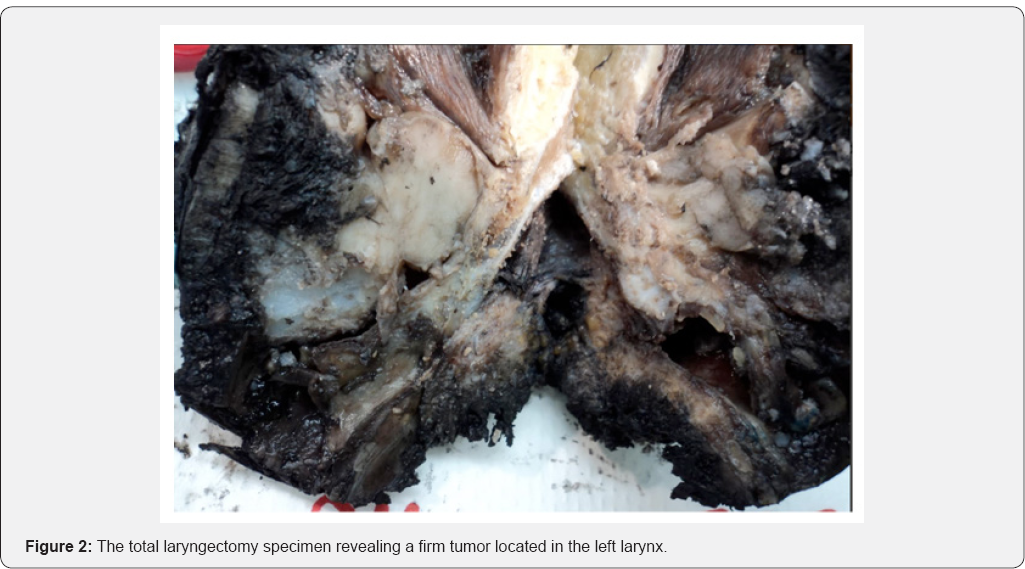
Within the total laryngectomy specimen, a firm tumor located in the left larynx was identified (Figure 2).The
tumor extended proximally from the false vocal cords, and distally,
into the subglottis. The tumor invaded both, the thyroid and cricoid
cartilages. The resection superior and inferior margins were negative.
The anterior margins were 4mm, and the posterolateral and posterior
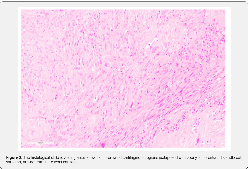
margins appeared to potentially involve the tumor. The histological
findings, shown in Figure 3,
revealed areas of well-differentiated cartilaginous regionsjuxtaposed
with poorly- differentiated spindle cell sarcoma, arising from the
cricoid cartilage. This finding was consistent with dedifferentiated
chondrosarcoma.


A PET scan was completed to rule out persistent or
recurrent disease in the pharyngeal region. There was no evidence of
hypermetabolic adenopathy, pulmonary nodules, or hypermetabolic activity
in the neckintraabdominal or pelvic organs. However, focal
hypermetabolism was present in the superior aspect of thesternal body,
either associated with a fracture or alternatively, a metastatic lesion.
Further, imaging with theMRI revealed a healing non-displaced sternal
fracture. There was no evidence of metastatic bone disease.
Radiation treatment to the laryngeal bed is
recommended for intermediate and poorly dedifferentiated chondrosarcoma
due to an ominous prognosis, high risk of local recurrence and distant
metastases. Furthermore, in the patient's case, radiation is recommended
due to the close margins revealed in the total laryngectomy specimen.
In this case, a total dose of 66Gy in 33 fractions of external beam
radiation therapy was completed. The patient will continue to receive
ongoing assessment, including 3-monthly appointments for the first two
years, 4-monthly appointments in the third year, and following with
6-monthly appointments. Moreover, a chest x-ray twice annually, and
further CT scans as indicated.
Discussion
Laryngeal chondrosarcoma is the most common non- epithelial laryngeal neoplasm [7,13-15].
The incidence of chondrosarcoma increases with age, with the majority
of cases arising in the sixth and seventh decade of life [3,6,7,10,11,13,1517].
Unlike other head and neck chondrosarcomas, laryngeal chondrosarcomas
are relatively slow growing, low- grade neoplasms. Local excision is
curative in most cases. Laryngeal chondrosarcomas typically originate
from hyaline cartilage. Approximately 75% of cases arise from the
posterior lamina of the cricoid cartilage [3,4,6-8,10-12,15,16,18-22].
The rest originate from the thyroid, arytenoid, epiglottis and accessory
cartilages [3,6,12,15,16,18,20-22]. The exact pathogenesis of laryngeal chondrosarcomas remains unknown [2,3,6,7,11,1517,19].
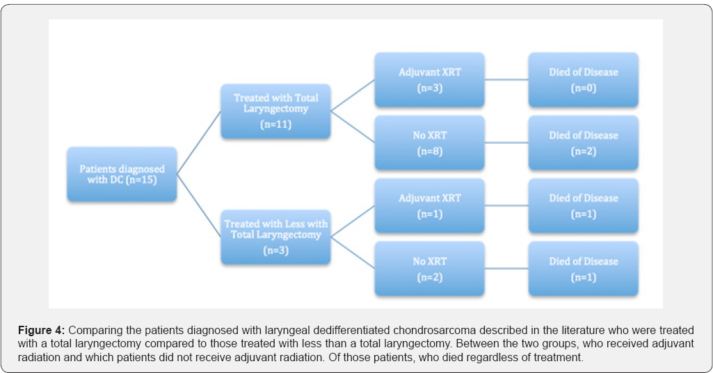
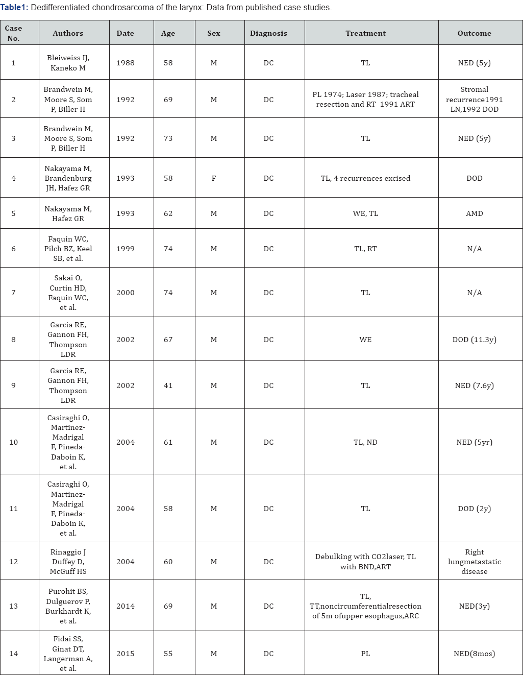
Dedifferentiated chondrosarcomas of the larynx are
extremely rare. Currently there are 14 cases reported in the literature,
including this report (Table 1 & Figure 4). The term dedifferentiated chondrosarcoma was first termed by Dahlin and Beabout in 1971 [9,18,22,23]. DC, a rare entity of chondrosarcoma, has an estimated incidence of 8-14% of all laryngeal chondrosarcomas [9]. DC are aggressive and present with a poor prognosis [13,20]. DC commonly arise among the older adult population, with a male predilection [10].
Compared to the cases of DC reported in the literature, the clinical
presentation, investigations and treatments were all similar.
Diagnosis of DC includes a combination of pertinent
information on history, physical examination, imaging, and histological
and immunocytochemical analyses. Definite diagnosis can be established
by incisional biopsy and histopathological examination. The utility of
FNA has only been described in 1 DC case. FNA can be performed in an
outpatient setting with minimal complications; it is also efficient and
cost- effective. Despite the advantages, the senior author does not
recommend FNA in the case of chondrosarcoma; there is a risk of
collecting non- representative tissue, it is difficult distinguishing
benign and malignant tumors, and grading and classifying lesions [9].


Symptomology
Presenting symptoms vary depending on the anatomic
location of the tumor. In the literature, common symptoms reported
include hoarseness due to narrowing of the glottis and compression of
the inferior laryngeal nerves, dyspnea and airway obstruction as a
result of endolaryngeal and subglottic growth, dysphagia due to
extralaryngeal growth of the tumor originating in the posterior cricoid,
and a painless neck mass from tumor the involving the thyroid cartilage
[7,10,11,12,13,18].
Rapid progression of symptoms over days or weeks should raise the
suspicion of the lesion may be more aggressive than the standard
laryngeal well-differentiated chondrosarcoma.
Histopathology
Histopathologic grading of chondrosarcomas is based
on the criteria first described by Lichtenstein and Jaffe in 1943. The
grading system stratifies the tumor into three different grades, from
low to high grade according to cellularity, nuclear size and
pleomorphism, necrosis and mitotic activity. This classification system
assists with delineating the tumor's aggressiveness and prognosis, and
subsequent treatment. Higher-grade neoplasms are associated with higher
aggressiveness and a poorer prognosis [6,7,15,16,20,23]. Histological analysis remains the gold- standard for diagnostic purposes [3,6,10].
The histopathologic hallmark of DC concluded in case studies is the
presence of an abrupt transition between low grade cartilaginous
component juxtaposed on a high- grade, noncartilaginous, spindle cell
sarcoma with pleomorphism, vesicular nuclei, giant cells, and numerous
mitoses[4,5,9,10,13,18,23]. The cartilaginous portion of the tumor can contain some cells of DC, and thus the entire tumor must be examined.
Morphologically, the features of DC are similar to
undifferentiated sarcoma, osteosarcoma, rhabdomyosarcoma, leiomyosarcoma
or angiosarcoma [6,10,13,18].
Immunohistochemistry
Immunochemical findings were not described in this
case, however other studies concluded that the malignant spindle cell
component of DC is strongly positive for vimentin, focally positive for
alpha-1-anti-chymotrypsin, and negative for cytokeratins, S-100 protein,
desmin and muscle-specific act in [9,13,18,22].
Imaging
According to the literature, CT scan is the imaging
modality of choice. Stippled to coarse calcifications within the tumor
is a patho gnomonic finding for laryngeal chondrosarcoma. This feature
can be seen in tumors of any grade. Thus, imaging alone is not
sufficient to make the diagnosis of dedifferentiated chondrosarcoma [2,
4,6,7,10,12,15,16,18]. This finding cannot be appreciated on MRI. MRI is
a useful complementary image modality for determining the extent of the
tumor, as well as treatment planning and prognosis.
Destructive invasion of the cartilage, bone or soft tissue may suggest a high-grade tumor [10].
One study outlined the feasibility of multimodality imaging with PET
and MRI, including DW-MRI. PET and MRI provide additional information
regarding the hyper metabolic, aggressive dedifferentiated component,
and the hyper metabolic, low-grade component [4].
Treatment
The standard treatment for laryngeal cartilaginous
tumors is surgical excision, with preservation of the function of the
larynx, if feasible [2-4,8,11,14,16,17,20]. However, due to the
aggressiveness of the DC, the definitive treatment remains total
laryngectomy [4,21].
Nonetheless, partial laryngectomy has been reported in a few cases. It
is also recommended that patients with laryngeal DC receive adjuvant
radiotherapy [2].
Although in this case, the patient received adjuvant radiation therapy,
there have been previously reported cases in the literature in which
the patient did not receive adjuvant therapy following surgical excision
[5,17]. The role of radiation therapy remains undetermined [4].
Follow-up reveals a variable clinical course among the reported cases.
Some patients remain disease free, whilst others develop metastatic
disease [10,20].
Prognosis
Compared to low-grade chondrosarcomas, DC has been
shown to have a poorer prognosis with a high rate of recurrence and a
predisposition of distant metastases [3,4,8,13,18,23].
The average life-expectancy reported is 6 months, and a 5-year survival
rate of 10.5%. Approximately 70% of patients develop
pulmonarymetastatic disease some time during their disease course [18,23].
Conclusion
DC is a rare entity in the larynx. DC typically
arises in the cricoid cartilage, presenting with rapidly Progressive
dyspnea, hoarseness, dysphagia, and a painless neck mass. Due to the
aggressiveness of thetumor, it is essential to include DC as a
differential diagnosis among the other laryngeal tumors [24,25].
Acknowledgement
Thank you to Dr. Brent Wilde for providing
photographs of the histology slide and of the gross specimen of the
dedifferentiated chondrosarcoma of the larynx.
For
more Open access journals please visit our site: Juniper Publishers
For
more articles please click on Journal of Cell Science & Molecular Biology


Comments
Post a Comment