Protein Expression Profile of Oral Premalignant Lesions (OPLs)- Juniper Publishers
Juniper Publishers- Journal of Cell Science
Abstract
The progression in cure, early detection and
degeneration of oral squamous cell carcinoma (OSCC) remain a key factor
to improve the survival rate of patients, in which an elevated
proportion of patients are diagnosed at an advanced stage. Current
developments in molecular biology research enhanced the understanding of
molecular process in OSCC progression and led to identification and
characterization of numerous biomarkers. These biomarkers are expected
to facilitate the early detection of primary and relapsed tumors. In the
present research, we evaluate potential biomarkers for the early
detection of OSCC.
Keywords: Carcinoma; Biomarkers; Protein; Serum; Cancer; Tumor
Introduction
Oral cancer is a subgroup of head and neck cancer
which affects various regions within oral cavity i.e., lips, tongue,
salivary glands, and gums. It is the sixth most common cancer,
approximately 3% of the total cancer burden and results in 128,000
annual deaths globally [1,2]. The most common type of oral cancer is
oral squamous cell carcinoma (OSCC), which accounts 90% of all oral
cancer cases. Patients with OSCC are often diagnosed at a late stage,
thus high recurrence rate occurs after treatment, especially in those
with neck lymph node metastasis [3]. Despite clinical and treatment
advances, the overall 5-year survival rates for oral cancer remains low
and stagnant during past few decades [4,5]. Although OSCC is commonly
diagnosed through oral examination followed by histopathology and
computed tomography/positron emission tomography scanning, there has
been continuous interest in developing serum protein biomarkers to aid
the diagnosis. Tumor antigens are promising diagnostic biomarkers for
human cancers showing the clinical utility of tumor antigens, such as
carcinoembryonic antigen (CEA), CA-50, CA19-9 and squamous cell
carcinoma antigen (SCCA) for OSCC detection [6-9].
Serum SCCA appeared to be more sensitive than the
other tumor antigens and positive in 38.1% and 41.4% of OSCC patients
under testing [8,10]. Studies also revealed other potential serum
protein biomarkers i.e., CYFRA 21-1 (cytokeratin 19-fragments), tumor
polypeptide antigen (TPA) and insulin-like growth factor binding protein
3 [9,11]. CYFRA 21-1 has been demonstrated
as biomarker for other solid tumors, whereas TPA is a serine protease
found in rapidly growing tissue due to its role in forming intermediate
filaments of the cellular cytoskeleton, making it a promising candidate
for cancer detection. Both CYFRA 21-1 and TPA levels were found to be
significantly higher in OSCC patients compared to healthy controls and
benign tumor patients, and both reduced in 2–3 weeks after surgical
resection of their OSCC lesions [11]. Although the testing serum protein
biomarkers as a simple diagnostic tool for oral/head and neck cancer
has been well demonstrated but still needs to be further validated in
large clinical trials. In this study, proteomics analysis of serum for
early detection, evaluation, aggressiveness and occurrence of OSCC were
summarized. The emphasis was placed on early detection by serum with
histological defined oral carcinoma patients. Protein in tissues, saliva
and serum may more accurately reflect the progression of OSCC, so novel
approach for the depth research strategy and the sample choice for
proteomics are of importance in OSCC biomarker discovery.
Material and Methods
Sample collection
Information about chewing habits and other
characteristics of the study participants was acquired. Those with the
habit were questioned for the frequency of the habit in number per day
and duration of the habit in years. The type of lesion in the cases was
decided on the basis of careful observation of the oral cavity and then
blood samples were collected from People’s College
of Dental Science and Research Centre, Bhanpur Bhopal, over a
period of one month. 2-3ml of blood were taken as samples from
the patients having oral premalignant lesions (OPL) (leukoplakia
and/or erythroplakia) and stored in sterile vials containing
1.5mg/ml EDTA and stored at -20 °C. Individuals who were not
addicted to smoking, tobacco or not habitual areca nut consumer
and were not having any prior history of cancer and who had
never smoked before in his or her lifetime are defined as a never
smoker and were considered as control.
Determination of Protein Content
Lowry method
The protein content of serum was determined 1mg/mL
stock solution of Bovine serum albumin (BSA) was prepared.
BSA was dissolved in 0.1 N NaOH and used as standard [12]. For
preparation of standard curve of BSA different concentration that
is 100μg/mL to 1000μg/mL of BSA was prepared in clean glass
tubes and volume was made up to 1mL using Millipore water.
To each tube, 5mL of alkaline solution was added and the tubes
were incubated at room temperature for 10min. After incubation,
0.5mL of Folin’s reagent was added and was incubated at room
temperature in dark for 30 minutes. Violet to blue colour was
developed. The intenity of colour was propotional to protein
concentration. After incubation the absorbance was measured
at 660nm. Standard curve was prepared by plotting protein
(BSA) concentration on X-axis and O.D on Y-axis. A blank was
prepared by taking 1mL of Millipore water in tube in which 5mL
of alkaline solution and 0.5mL of Folin’s reagent were added. All
the procedures were carried out in triplets. Estimation of protein
contents was done in a same manner as BSA standard curve.
Sodium dodecyl sulphate polyacrylamide gel electrophoresis (sds-page)
Poly acrylamide gel electrophoresis in the presence of
sodium dodecyl sulphate was performed according to LaemmLi
[13]. The SDS-PAGE was performed using a 12% separating
gel and 4% stacking gel. The samples were heated for 5min at
100 °C in capped vials with 1% (w/v) SDS in the presence of
β-mercaptoethanol. Electrophoresis was performed at a 125
V for 4h in Tris- HCl buffer of pH 8.3. After electrophoresis,
proteins in the separating gel were made visible by staining
with Coomassie Brilliant Blue R-250. The standards were used
to make a plot of log molecular weight versus mobility of the
protein band were myosin (200kDa), β-galactosidase (120kDa),
bovine serum albumin (91kDa), glutamate (62kDa), ovalbumin
(43kDa), glyceraldehyde-3-phosphate dehydrogenase (36kDa),
carbonic anhydrase (29kDa), myoglobin (26kDa), and lysozyme
(14kDa) as markers [6] (Table 1).

Results and Discussion
The OPLs (leukoplakia/erythroplakia) case sample was
collected from OPLs patients of PCDS&RC, People’s University,
Bhopal the sample size was 20 in which 3 were females and
17 were males under the age group of 18 to 60. Taking into
consideration the addictions, 11 of them were habitual of
smoking either bidi or cigarette, 6 were betel nut consumers
whereas most of them were pouch/gutkha consumers. Among
the sample population, female: male ratio observed was 3:17,
where females account for 30% and males 70%. Total population
(OPLs cases) age range varies from 18 to 68 years, of which 18-
68 and 25-54 was age range of males and females respectively.
Among the observed population 38% were smokers, 21% were
betel nut consumers and majority i.e., 41% were pouch and
gutkha consumers. Biomarkers have been wide accepted in
other disciplines but there is no consensus for their use in oral
malignancies. Despite recent advances in surgical, radiotherapy,
and chemotherapy treatment protocols, the survival of patients
with OSCC still lacks significant improvement. This unsatisfactory
treatment may be explained by the fact that OSCCs frequently
present with extensive local invasion and advanced stages
[14,15]. That makes necessary the development of new tools for
the diagnosis and prognosis.
Protein Profiling
Total protein
From the known concentration of protein, the standard
curve was plotted, and protein content of the clinical isolates
were observed. The total proteins were estimated which
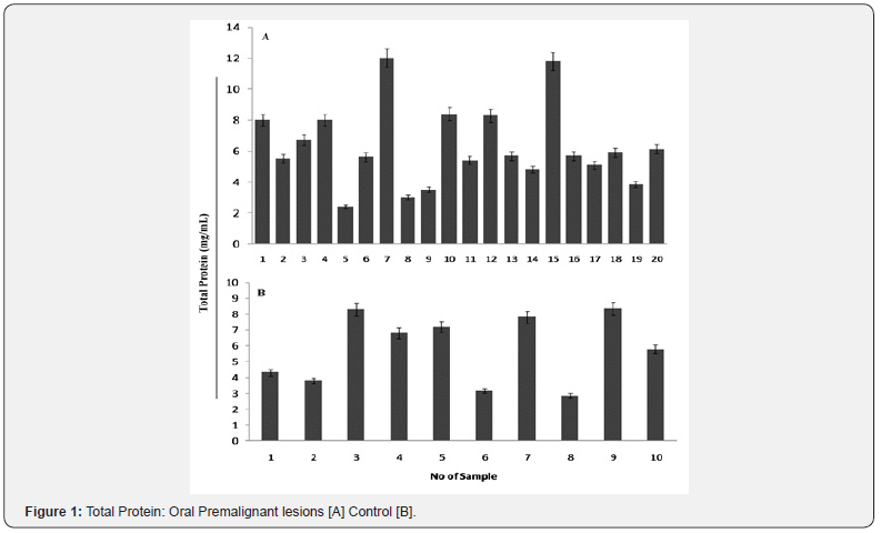
revealed the difference in Oral premalignant lesions. The protein
concentration ranged from 3.2-11.8mg/mL. Control group
the protein concentration ranged from 2.87-8.35 (Figure 1).
Oxidation of protein plays an important role in pathogenesis of
cancer and studies have demonstrated decreased protein levels
in cases of OPMD’s and oral malignancy [16,17]. In oral cancer,
tobacco and areca nut related habit leading to tissue damage and
resultant free radicals play a major role as an aetiologic factor.
These habits are seen commonly in all the ages and both the sex. The serum protein levels were decreased in OSMF, OL and NS
but increased in OM. This difference was statistically significant.
These findings are fill agreement of with the findings of Patidar
et al. [18] and Rajendran et al. [19] in OSMF participants and
Dawood et al. [20] in OM participants. In contrast our results did
not simulate with the finding of Chandran et al. [16] in OM group
where the plasma protein levels were found to be decreased. The
increase in serum protein levels may be explained in terms of
inflammatory reaction associated with oral malignancy.

Sodium dodecyl sulfate polyacrylamide gel electrophoresis
SDS-PAGE analysis of various samples revealed at least 06
types of banding patterns, with the number of bands ranging
from 25 to 40. The maximum number of sample 26.6%) had
a banding pattern with 04 bands. Proteins with molecular
weights 66kDa, 75.0kDa, 95,110 and 140kDa were consistently
present in the in the pattern. The pattern 2 showed number of
16.6 % sample had a banding pattern with 03 proteins bands
with molecular weights 11.5,23, and 90.5kDa. The pattern
3 (10% sample) showed 04 proteins bands with molecular
weights 118.5, 23, 66 and 97kDa. Pattern 4 showed number of
23.3 % sample had a banding pattern with 04 proteins bands
with molecular weights 14, 18, 42 and130kDa. Pattern 05 also
showed number of 10 % sample with 04 proteins bands with
molecular weights 67.5,75,95 and110kDa. Pattern 6 revealed
03banding pattern with molecular weights14.5, 38.5, 55.5kDa.
Feng et al. [21] measured the level of some biomarkers (SCCA,
Cyfra 21-1, epidermal growth factor receptor (EGFR) and Cyclin
D1) in an attempt to determine the usefulness of their combined
determination in the diagnosis of OSCC. They concluded that
Cyclin D1, the product of the CCND1 gene located on chromosome
11q13, had the highest diagnostic specificity. Moreover, the
combined detection of EGFR and Cyclin D1 (36kDa) had the
highest sensitivity, specificity and accuracy. A previous study
Capaccio et al. [22] demonstrates that Cyclin D1expression was
significantly associated with the presence of occult lymph node
metastases. These data suggest that the immunohistochemical
analysis of Cyclin D1 expression in diagnostic biopsy samples
may be an additional tool for selecting patients to be treated
with elective neck dissection.
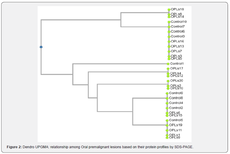
The dendrogram showed that the samples [12] were grouped
in two closely related clusters. The clusters of Oral Premalignant
lesions were significantly different from an unrelated to that of
the control. It was also seen that sample from control tended
to fall close together on cluster analysis. In our study, the SDSPAGE
pattern revealed several characteristic bands common to
all samples.
The sample under study was divided in to 2 clusters with
cluster 1 having 12 sample and cluster 2 with 18 samples
having a similarity 82.5% and dissimilarity of 17.5% in the
jaccard’s coefficient scale ion the dendogram (Figure 2). The
cluster 1 was further divided into 1a and 1b with 3(OPLs-18,
OPLs-08 OPLs-14,) and 9 (Control-10, Control-07, Control-6,
Control-3, OPLs-16, OPLs-13, OPLs-07, OPLs-03, OPLs-05)
sample respectively. The cluster 2 was also divided in to two sub-cluster 2a (1) (Control-1) and 2b (17) (OPLs-17, OPLs-04,
OPLs-12, OPLs-20, OPLs-02, OPLs-10, Control-09, Control-08,
Control-04, Control-02, OPLs-06, OPLs-15, Control-5, OPLs-
19, OPLs-11, OPLs-01, OPLs-09). Cluster 2a having 1 sample
showed similarity 88.7% and dissimilarity of 10.5%. 2b showed
similarity 87.3% and dissimilarity of 12.3% (Figure 2).

The sub-cluster 2(b) was also sub divided into sub sub cluster
as 2b1 (OPLs-17, OPLs-04, OPLs-12) showed similarity 87.9%
and dissimilarity of 12.1%. Cluster as 2bII (OPLs-20, OPLs-02,
OPLs-10, Control-09, Control-08, Control-04, Control-02, OPLs-
06, OPLs-15, Control-5, OPLs-19, OPLs-11, OPLs-01) showed
similarity 86.1% and dissimilarity of 13.9%. These differences
between the samples are due to the difference in their protein
profile which can be mediated due to the difference in the oral
premalignant lesions therapy. The study clearly indicates that
the profile of total protein from Oral Premalignant lesions
can be used for developing classification pattern. The cluster
2bII was also sub divided into sub cluster as 2b1Ia and 2b1Ib
(Control-09, Control-08, Control-04, Control-02, OPLs-06, OPLs-
15, Control-5, OPLs-19, OPLs-11, OPLs-01) showed similarity
87.9% and dissimilarity of 12.1% (Figure 2). Cluster analysis has
been used, allowing one to make a more objective interpretation
of immunoprofiles, based on staining with multiple antibodies,
and holding great promise for the immunohistochemical
classification of tumors [23].
Ideally, a good clinical test requires high sensitivity and
specificity. The oral cavity is commonly subject to inflammation
from a variety of causes, including trauma, dental plaque,
infection and certain mucocutaneous inflammatory diseases.
Whether such oral inflammation (non-neoplastic conditions)
affects the levels of the potential OSCC serum biomarkers is
essentially unknown, because most studies investigated the
potential biomarker levels only in OSCC and non-OSCC, without
regard to other inflammatory conditions present [24]. If any OSCC
biomarkers levels increases in the presence of oral inflammation
to the level of OSCC patients, it would result in a high false
positive rate and greatly reduce the value of that biomarker
in clinical use for detection. Many of reported potential OSCC
biomarkers, such as IL-6 [25], IL-8, IL-1 β [26], basic fibroblast
growth factor [27] and molecules related to oxidative stress [27]
are known to be important factors involved in inflammation
and/or wound healing [28]. Indeed, the levels of some of these
constituents have been reported to be significantly higher or
lower in periodontitis or OLP patients who did not have OSCC
[29]. Therefore, research that validates any potential OSCC
biomarker with individuals having common non-neoplastic oral
inflammatory diseases is necessary in order to establish the
reliability of that salivary OSCC biomarker
Conclusion
Serum biomarkers obtained represent a promising approach
for oral cancer detection, and an area of strong research interest. However, some issues/challenges needs to be determined in
order to establish this approach as a reliable, highly sensitive and
specific for clinical use, including lack of consistency of serum
sample collection, processing and storage; wide variability in
the levels of potential oral carcinoma serum biomarkers in both
non-cancerous individuals and oral carcinoma patients; and
further validation of oral carcinoma serum biomarkers with
individuals either a chronic oral inflammatory disease or other
types of cancers, but do not have oral carcinoma. Research for
eventual standardization especially biological and physiological
variance affecting the potential biomarkers gained importance
in serum diagnostics. This approach can be useful in monitoring
non-cancerous disease activity applying serum biomarkers for
other forms of cancer.
Acknowledgement
The authors are thankful to People’s University, Peoples
Group, Bhopal, for laboratory facilities, to carry out this research
work.


Comments
Post a Comment